First of all we need to know some terms related to conjugation.
Conjugation: conjugation is the process by which one bacterium transfers genetic material to another through direct contact. During conjugation, one bacterium serves as the donor of the genetic material, and the other serves as the recipient. The donor bacterium carries a DNA sequence called the fertility factor, or F-factor.
Conjugants: Conjugant is a member of a mating pair of organisms or gametes undergoing conjugation.
Micronuclei: Micronuclei are extra-nuclear bodies that contain damaged chromosome fragments and/or whole chromosomes that were not incorporated into the nucleus after cell division.
Macronuclei: A macronucleus is the larger type of nucleus in ciliates. It controls the non-productive cell functions, such as metabolism.
Process of conjugation of paramecium is simply discussed below:
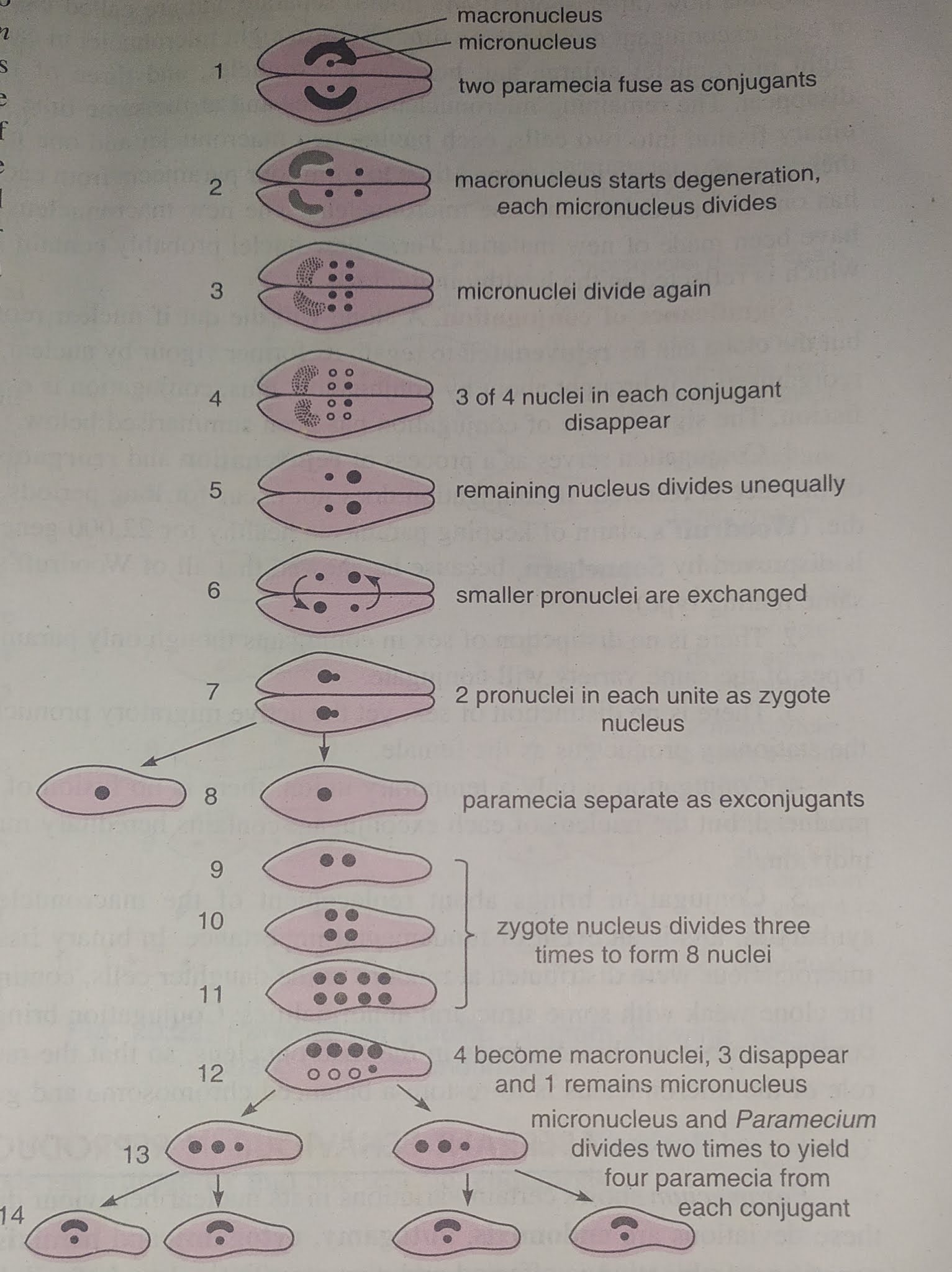
- Firstly two pre-conjugants of opposite matting types come together adhere.
- They stop feeding, and their oral groove apartures disappear.
- Then a protoplasmic bridge formed between them and these individuals are now called conjugants.
- The macronucleus begins to disintegrated and finally disappeared.
- The micro-nucleus of each individuals divides two times. One of them has a reductional division. So, a haploid micro-nucleus are produced in each conjugant.
- 3 of these 4 micro-nucleus disappeared in each. only one remain, and this nucleus divides mitotically and forms two unequal pro-nucleus .
- One of them pro-nucleus of each conjugants crosses over the protoplasmic bridge and fuse with the other pro-nucleus of conjugant forming a synkaryon.
- After about 12-48 hours conjugants are separate now and called ex-conjugants.
- The synkaryon of each ex-conjugants divides 3 times to form 8 nuclei. 4 becomes macronucleus, 3 disappeared, and one remains micro nucleus.
- The remaining one divides and at the same time ex-conjugants divides by binary fission into two cells.
- Each have two macro and one micro nucleus.
- The cell and their micro-nuclei divides second time to form a paramecia, so that each has one macro and one micro-nucleus.

